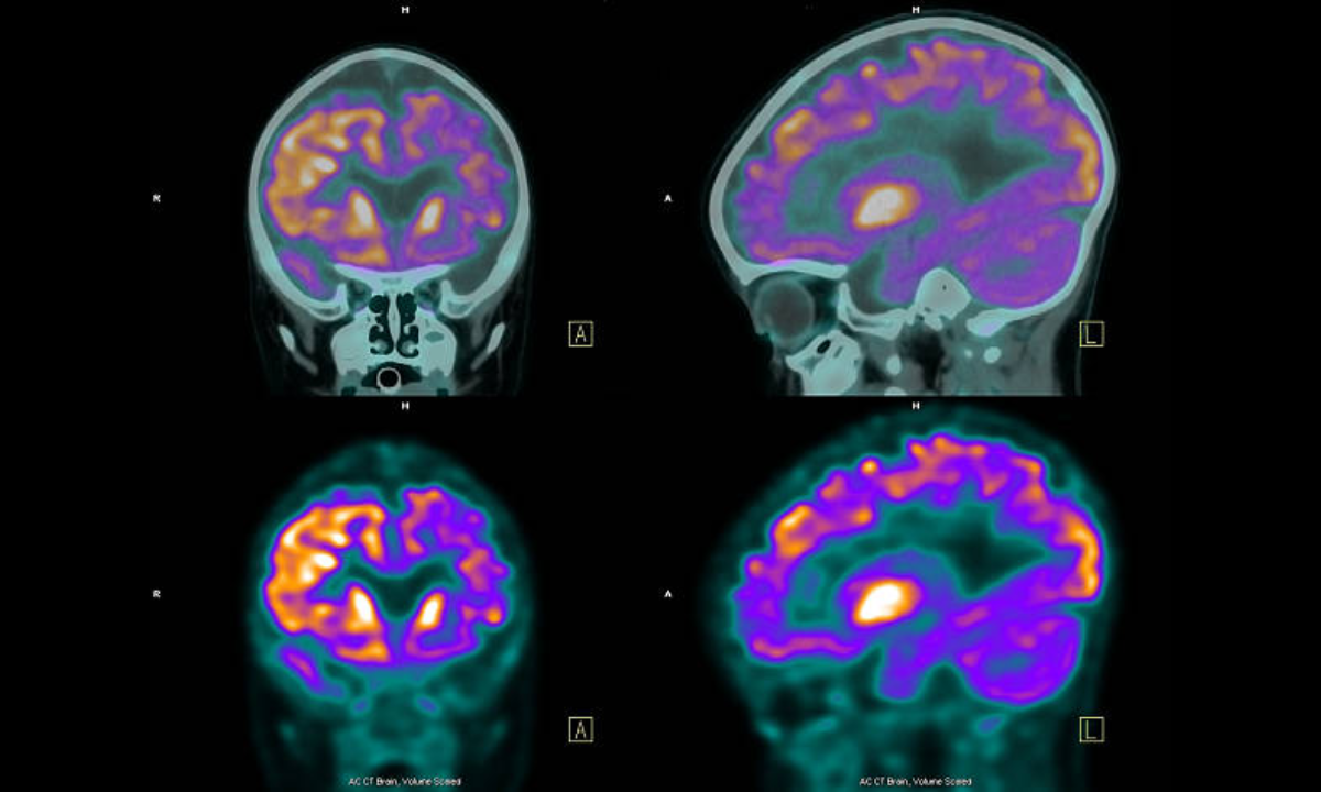
A Positron Emission Tomography scan, commonly known as a PET scan, is an advanced imaging technique that helps evaluate brain function at a cellular level. Unlike MRI or CT scans, which primarily show structural details, a PET scan reveals how brain tissues are working by measuring metabolic activity and blood flow. In neurology, this test plays an important role in assessing complex brain disorders where structural imaging alone may not provide complete answers.
During the procedure, a small amount of a safe radioactive tracer is injected into the bloodstream. This tracer travels to the brain and highlights areas of increased or decreased metabolic activity. The scan then produces detailed images that help identify abnormal patterns. PET scans are especially valuable in diagnosing and monitoring conditions such as epilepsy, brain tumors, dementia, and certain movement disorders. When recommended appropriately, it provides critical insights that guide accurate diagnosis and personalized treatment planning.
A PET scan is typically advised when neurological symptoms suggest underlying brain dysfunction that cannot be fully explained by routine imaging. It may be recommended for patients with unexplained seizures, progressive memory decline, suspected brain tumors, or movement abnormalities.
In conditions like epilepsy, PET scans help locate the specific region of the brain responsible for seizure activity. In cases of cognitive decline, such as suspected Alzheimer’s disease, the scan can detect characteristic patterns of reduced metabolism in certain brain regions. For patients with suspected brain tumors, PET imaging helps differentiate between active tumor tissue and scar tissue, aiding in treatment decisions. The test is usually performed when clinical evaluation and other imaging results require further clarification.
Neurological symptoms that may lead to a PET scan recommendation include persistent or unexplained seizures, memory loss, confusion, personality changes, difficulty speaking, or progressive weakness. Patients with movement-related symptoms such as tremors or stiffness may also require PET imaging when disorders like Parkinson’s disease are suspected.
In individuals with suspected brain tumors, symptoms may include persistent headaches, vomiting, vision changes, or new neurological deficits. A PET scan helps determine the underlying cause of these symptoms by identifying abnormal metabolic patterns within the brain.
PET scanning is often part of a comprehensive neurological evaluation. Before recommending the test, a detailed clinical examination is performed along with other imaging studies such as MRI or CT scans. In some cases, PET may be combined with CT or MRI to provide both structural and functional information in a single session.
The procedure itself is safe and typically completed within a few hours. After injection of the tracer, a waiting period allows it to distribute throughout the body. The patient then lies comfortably on the scanning table while the machine captures images. The results are carefully interpreted by specialists to identify areas of abnormal brain metabolism, which helps confirm or refine the diagnosis.
While a PET scan is not a treatment, it plays a crucial role in guiding treatment decisions. In epilepsy, it helps identify candidates for surgical intervention by accurately locating seizure focus areas. In dementia, it assists in distinguishing between different types, ensuring appropriate medical therapy and long-term planning.
For brain tumors, PET imaging helps determine whether a lesion is active and aggressive, guiding surgery, radiation therapy, or chemotherapy planning. In movement disorders, it may support decisions regarding medication adjustments or advanced therapies. The information gained from PET imaging allows for more precise and individualized neurological care.
After a PET scan, patients can typically resume normal activities immediately. Drinking plenty of fluids helps flush the tracer from the body more quickly. The radioactive material used is minimal and naturally eliminated over time.
Follow-up care depends on the underlying neurological condition. Once results are available, a detailed discussion is conducted to explain the findings and outline the next steps. Ongoing monitoring, medication adjustments, rehabilitation, or further investigations may be recommended based on the diagnosis.
PET scans are generally safe and well tolerated. The amount of radiation exposure is low and considered medically acceptable when the test is clinically indicated. Allergic reactions to the tracer are extremely rare.
Pregnant or breastfeeding women should inform their doctor before undergoing the procedure, as special precautions may be necessary. Mild discomfort at the injection site can occasionally occur, but serious complications are uncommon. When performed under expert supervision, PET scanning remains a safe and reliable diagnostic tool.
You should consult a neurologist if you experience persistent seizures, progressive memory problems, unexplained headaches, movement difficulties, or sudden changes in behavior or cognitive function. Early evaluation is particularly important in conditions like suspected Alzheimer’s disease or seizure disorders, where timely diagnosis significantly influences treatment outcomes.
A PET scan is recommended only when clinically appropriate and after careful assessment. Under the guidance of Dr. Sudheer Pachipala, patients receive a thorough evaluation to determine whether PET imaging is necessary as part of a comprehensive neurological care plan.
Chat With Me