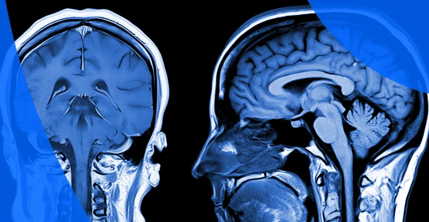
Magnetic Resonance Imaging (MRI) and Computed Tomography (CT) scans of the brain and spine are advanced imaging tools that help detect structural and functional abnormalities of the nervous system. Proper interpretation of these scans is crucial for accurate diagnosis and effective treatment planning. While the imaging technology captures detailed pictures, understanding what those images mean in relation to a patient’s symptoms requires expert neurological evaluation. A neurologist carefully correlates scan findings with clinical history, physical examination, and laboratory results to arrive at a clear and reliable diagnosis. Accurate MRI and CT scan interpretation plays a vital role in identifying conditions affecting the brain, spinal cord, nerves, and surrounding structures.
Brain and spine scans are typically advised when there is suspicion of underlying neurological disorders. These may include stroke, brain tumors, infections, inflammation, multiple sclerosis, epilepsy-related structural changes, head injury, spinal disc problems, spinal cord compression, congenital abnormalities, or degenerative spine disease. MRI is especially useful for evaluating soft tissues, including the brain, spinal cord, and intervertebral discs, whereas CT scans are often preferred in emergency settings such as trauma or suspected bleeding. The need for imaging arises when symptoms suggest structural or vascular causes that require visualization for confirmation.
Patients are usually referred for MRI or CT scans when experiencing persistent headaches, seizures, dizziness, weakness in the limbs, numbness, vision changes, speech difficulties, memory issues, back pain, neck pain, radiating limb pain, or difficulty walking. Sudden symptoms such as severe headache, loss of consciousness, or signs of stroke demand urgent imaging. Chronic symptoms like progressive weakness, balance problems, or ongoing spinal pain may also require detailed imaging to determine the underlying cause. Interpreting scan findings in the context of these symptoms ensures that treatment is tailored precisely to the patient’s condition.
Accurate diagnosis depends not only on obtaining high-quality imaging but also on expert interpretation. MRI and CT scans can reveal abnormalities such as tumors, hemorrhage, infarcts, demyelinating lesions, spinal disc herniation, spinal stenosis, fractures, infections, or degenerative changes. However, not all findings are clinically significant. Some changes may be age-related or incidental. A neurologist carefully distinguishes between significant pathology and harmless variations, preventing unnecessary anxiety or treatment. In certain cases, additional tests such as contrast imaging, MR angiography, nerve conduction studies, or laboratory investigations may be recommended to clarify findings and confirm the diagnosis.
Treatment depends entirely on the underlying condition identified on imaging. Management may include medications for stroke prevention, epilepsy control, inflammation, or infection. Structural issues such as disc prolapse or spinal cord compression may require physiotherapy, pain management strategies, or referral for neurosurgical evaluation. Brain tumors or vascular abnormalities may require multidisciplinary care involving neurologists, neurosurgeons, and oncologists. Early and precise interpretation of MRI or CT findings allows for timely intervention, which can significantly improve outcomes and reduce complications.
Follow-up imaging may be necessary to monitor disease progression or response to treatment. For example, patients with multiple sclerosis, brain tumors, or postoperative spine conditions often require periodic MRI scans to assess stability or improvement. Ongoing neurological assessment ensures that treatment remains appropriate and effective. Lifestyle modifications, rehabilitation programs, and medication adherence also play a key role in long-term recovery and prevention of recurrence.
MRI scans are generally safe, as they do not use radiation, but they may not be suitable for patients with certain implanted metallic devices. CT scans involve controlled radiation exposure, which is considered safe when medically indicated. Contrast agents used in some scans may rarely cause allergic reactions or kidney-related concerns, particularly in patients with pre-existing kidney disease. From a diagnostic perspective, misinterpretation or overinterpretation of imaging can lead to unnecessary procedures or delayed treatment. Therefore, specialist neurological review is essential for accurate clinical correlation.
You should consult a neurologist if you experience persistent or worsening headaches, seizures, unexplained weakness, sensory changes, chronic back or neck pain, balance disturbances, or any sudden neurological symptoms. Early evaluation and timely imaging can help detect serious conditions at an early stage. If you have already undergone an MRI or CT scan and require a detailed explanation of the findings, a specialist consultation ensures clarity, accurate diagnosis, and a structured treatment plan.
Comprehensive interpretation of MRI and CT brain and spine scans provides critical insight into neurological health. With expert evaluation, patients receive clear guidance, precise diagnosis, and personalized treatment strategies designed to protect and restore nervous system function.
Chat With Me