A CT Brain, also known as a computed tomography scan of the brain, is an advanced imaging test that uses specialized X-ray technology to create detailed cross-sectional images of the brain and skull. This diagnostic tool plays a crucial role in emergency and routine neurological care, allowing rapid assessment of brain structure, bleeding, swelling, tumors, stroke, fractures, and other abnormalities.
CT Brain scans are widely used because they are quick, non-invasive, and highly effective in identifying life-threatening conditions. In emergency situations such as head injury or suspected stroke, a CT scan often provides immediate and vital information that guides urgent treatment decisions. The procedure may be performed with or without contrast dye, depending on the clinical requirement.
A CT Brain scan is not a condition itself but an investigation recommended when certain neurological symptoms or concerns arise. It is commonly advised following head trauma, road traffic accidents, sudden severe headaches, unexplained seizures, weakness of limbs, loss of consciousness, or suspected stroke.
Doctors may also recommend a CT Brain to evaluate persistent headaches, brain infections, tumors, congenital abnormalities, hydrocephalus, or bleeding inside the brain. In some cases, it is used to monitor known neurological disorders or assess the outcome after brain surgery.
Patients are typically referred for a CT Brain when they experience symptoms that suggest a possible brain disorder. These may include sudden weakness or numbness on one side of the body, difficulty speaking, confusion, loss of balance, visual disturbances, repeated vomiting, or seizures.
Severe or unusual headaches, especially when accompanied by fever, neck stiffness, or altered consciousness, may also require imaging. In trauma cases, symptoms such as drowsiness, memory loss, persistent headache, or bleeding from the ear or nose are strong indicators for urgent scanning.
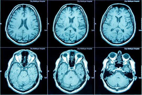
During the CT Brain procedure, the patient lies comfortably on a motorized table that slides into a circular scanner. The machine rotates around the head and captures multiple images, which are then processed by a computer to produce detailed brain images. The scan is painless and typically completed within a few minutes.
If contrast dye is required, it is administered intravenously to enhance visualization of blood vessels, tumors, or infections. A neurologist carefully reviews the images to identify abnormalities such as hemorrhage, infarction (stroke), mass lesions, swelling, fractures, or structural changes. CT Brain is particularly valuable in detecting acute bleeding and early stroke changes.
The CT Brain scan itself is a diagnostic tool and does not provide treatment. However, the findings play a critical role in guiding appropriate medical or surgical management. For example, if the scan reveals a stroke, immediate treatment such as clot-busting medication or other stroke interventions may be initiated. If bleeding is detected, urgent neurosurgical evaluation may be required.
In cases of tumors, infections, or structural abnormalities, the scan helps determine whether medications, surgery, or further imaging such as MRI is necessary. Accurate imaging allows for precise and timely treatment planning.
After a CT Brain scan without contrast, patients can usually resume normal activities immediately. If contrast dye was used, adequate hydration is recommended to help flush the dye from the body. Patients are generally observed briefly if they have received contrast, especially if they have a history of allergies or kidney concerns.
Follow-up care depends entirely on the scan findings. Your neurologist will explain the results clearly and outline the next steps, whether that involves medication, additional tests, referral to a specialist, or lifestyle modifications.
CT Brain scans are considered safe and widely performed. However, as with any imaging involving radiation, there is minimal exposure to X-rays. The radiation dose is carefully controlled and kept as low as possible while ensuring high-quality images.
When contrast dye is used, there is a small risk of allergic reaction, ranging from mild itching to rare severe reactions. Patients with kidney disease may require special precautions before receiving contrast. Your doctor will assess individual risk factors before recommending the procedure.
Immediate medical attention is necessary if you experience sudden weakness, difficulty speaking, severe headache unlike any previous one, seizures, confusion, or head injury with loss of consciousness. Early evaluation and imaging can be lifesaving.
If you have persistent neurological symptoms such as chronic headaches, unexplained dizziness, or changes in memory or behavior, consultation with a neurologist is advisable. Timely assessment with appropriate imaging such as a CT Brain scan ensures accurate diagnosis and early intervention, leading to better outcomes and peace of mind.
Chat With Me