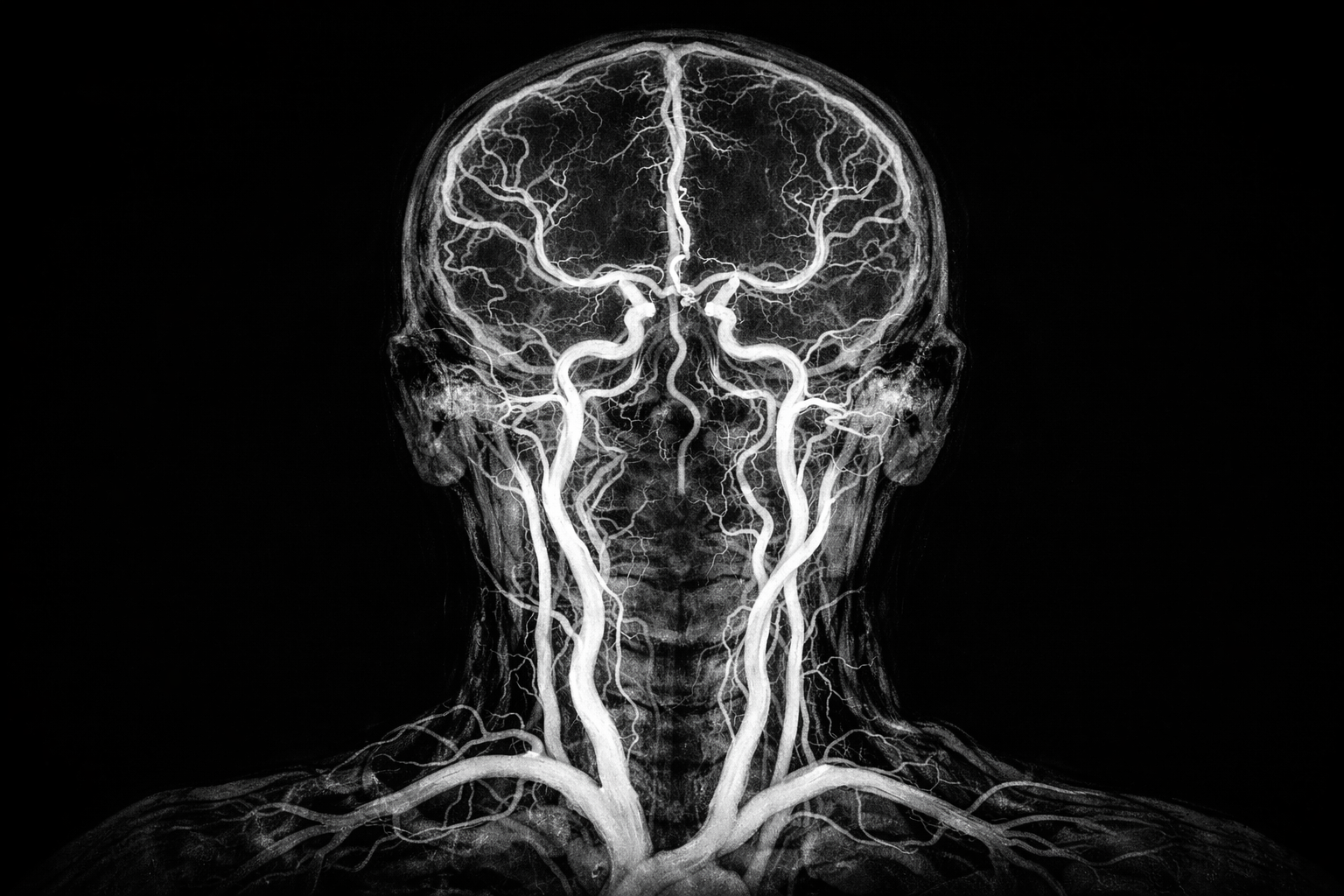
MR Angiography (MRA) and CT Angiography (CTA) are advanced imaging techniques used to visualize blood vessels in the brain, neck, and other parts of the body. These tests help assess the structure and flow of blood through arteries and veins, allowing neurologists to detect narrowing, blockages, abnormal vessel formations, or aneurysms. MRA uses magnetic resonance imaging and does not rely on ionizing radiation, while CTA uses computed tomography combined with contrast dye to produce highly detailed images. Both tests are non-invasive, precise, and play a critical role in diagnosing vascular conditions affecting the nervous system.
MR Angiography and CT Angiography are typically advised when there is suspicion of blood vessel abnormalities that may affect brain function or increase the risk of stroke. Common reasons include atherosclerosis (hardening or narrowing of arteries), aneurysms, vascular malformations, arterial dissections, or reduced blood flow to specific brain regions. These conditions may develop due to factors such as high blood pressure, diabetes, high cholesterol, smoking, genetic predisposition, or age-related vascular changes.
Patients who require angiographic imaging may present with symptoms such as sudden or severe headaches, unexplained dizziness, weakness or numbness on one side of the body, vision problems, difficulty speaking, or episodes suggestive of transient ischemic attacks or stroke. In some cases, these tests are recommended even without prominent symptoms, especially in individuals with known risk factors or previous vascular events, to monitor disease progression or treatment response.
During MR Angiography or CT Angiography, detailed images of blood vessels are obtained after administering a contrast agent that enhances vessel visibility. The scans allow the neurologist to assess vessel size, shape, and blood flow patterns with high accuracy. These findings help confirm or rule out conditions such as aneurysms, blockages, stenosis, or abnormal vessel connections. The choice between MRA and CTA depends on the clinical situation, patient factors, and the level of detail required.
Treatment decisions are guided by the angiography results and the underlying condition identified. Some vascular abnormalities may be managed with medications to control blood pressure, cholesterol, or clot formation. In more serious cases, interventional procedures such as endovascular treatment, stenting, or surgical correction may be recommended. Angiographic imaging is essential in planning these treatments and ensuring they are performed safely and effectively.
After treatment, follow-up imaging with MR Angiography or CT Angiography may be advised to monitor blood vessel healing and ensure adequate blood flow. Patients are often guided on lifestyle modifications, medication adherence, and regular neurological follow-up to reduce the risk of recurrence. Ongoing monitoring helps detect any new or progressing vascular changes at an early stage.
Both MR Angiography and CT Angiography are generally safe procedures. CTA involves exposure to a small amount of radiation, and both tests may require contrast dye, which can rarely cause allergic reactions or kidney-related issues in susceptible individuals. These risks are carefully assessed beforehand, and appropriate precautions are taken to ensure patient safety.
A neurologist should be consulted promptly if there are symptoms such as sudden weakness, speech difficulty, severe headache, vision loss, or repeated episodes of dizziness. Individuals with known vascular risk factors or a family history of aneurysms or stroke may also benefit from evaluation even in the absence of symptoms. Early assessment and timely imaging play a crucial role in preventing serious neurological complications and ensuring optimal long-term outcomes.
Chat With Me