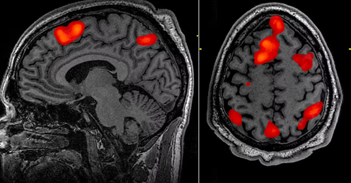
Functional MRI (fMRI) is an advanced, non-invasive brain imaging technique that measures and maps brain activity. Unlike a conventional MRI scan, which provides detailed images of brain structure, functional MRI evaluates how different areas of the brain function in real time. It works by detecting subtle changes in blood flow and oxygen levels that occur when specific parts of the brain are active. When a person performs a task such as speaking, moving, thinking, or responding to visual stimuli, the involved brain regions require more oxygen, and this change can be captured and analyzed.
In modern neurological practice, fMRI plays an important role in understanding complex brain disorders and in planning treatments, particularly before brain surgery. It helps neurologists and neurosurgeons identify critical areas responsible for speech, memory, movement, and sensation, ensuring safer and more precise medical interventions.
Functional MRI is not a treatment for a disease itself, but rather a diagnostic and planning tool used to evaluate conditions that affect brain function. It is commonly recommended for patients with brain tumors, epilepsy, stroke, traumatic brain injury, neurodegenerative disorders, and certain psychiatric conditions. In patients being evaluated for brain surgery, especially for tumors or seizure disorders, fMRI helps identify essential functional areas to minimize the risk of post-operative deficits.
It may also be advised when patients experience unexplained neurological symptoms such as persistent weakness, speech difficulty, memory impairment, or changes in behavior that require detailed functional brain assessment.
Patients who may benefit from a functional MRI often present with symptoms related to altered brain activity. These can include seizures, recurrent headaches with neurological deficits, speech disturbances, limb weakness, sensory changes, visual disturbances, balance problems, or cognitive decline. In some cases, individuals with suspected brain lesions may not have obvious symptoms, and fMRI is performed as part of a comprehensive evaluation before surgical intervention.
The test itself does not cause symptoms, and it is performed in a controlled clinical environment under the supervision of trained professionals.
Functional MRI is performed in a specialized MRI suite. During the procedure, the patient lies comfortably inside the scanner while performing specific tasks such as moving a hand, reading words, answering simple questions, or observing visual patterns. These tasks are carefully designed to activate particular regions of the brain. The scanner then records changes in blood oxygen levels, which are processed into detailed functional maps.
The data obtained from fMRI are combined with structural MRI images to provide a comprehensive understanding of both brain anatomy and function. This integration is particularly valuable in pre-surgical planning, epilepsy mapping, and research-based neurological assessments. The procedure is painless and typically takes between 30 to 60 minutes, depending on the complexity of the study.
Functional MRI itself is not a treatment modality, but it significantly influences treatment planning. In patients with brain tumors, it helps guide neurosurgeons in preserving vital areas responsible for speech and movement. In epilepsy management, fMRI assists in identifying seizure focus regions and determining surgical candidacy.
For patients with stroke or traumatic brain injury, the results may guide rehabilitation strategies by identifying preserved and affected brain regions. In certain neuropsychiatric disorders, fMRI findings can support tailored therapeutic approaches and advanced treatment planning.
Since fMRI is a non-invasive diagnostic procedure, no special recovery period is required. Patients can resume normal activities immediately after the scan. If the imaging was performed as part of surgical planning or evaluation of a neurological disorder, follow-up consultations are scheduled to discuss the results and determine the next steps in management.
When fMRI is used for pre-surgical mapping, post-operative care depends on the underlying condition and the type of surgery performed. Ongoing neurological monitoring and rehabilitation may be recommended when necessary.
Functional MRI is considered a safe procedure because it does not involve radiation exposure. However, as with any MRI scan, it may not be suitable for patients with certain implanted metallic devices, pacemakers, or severe claustrophobia. In rare cases, patients may experience mild discomfort due to lying still for an extended period.
If contrast material is used alongside structural imaging, there is a minimal risk of allergic reaction, although this is uncommon. A thorough medical history is always reviewed before the procedure to ensure safety.
You should consult a neurologist if you experience persistent neurological symptoms such as unexplained seizures, speech difficulties, limb weakness, memory problems, severe headaches, or sudden changes in behavior or cognition. Early evaluation is especially important when symptoms are progressive or worsening.
Functional MRI may be recommended when detailed assessment of brain function is necessary for diagnosis, surgical planning, or advanced neurological care. A timely consultation ensures accurate diagnosis, appropriate treatment planning, and better long-term outcomes.
Chat With Me